From Cath Lab to PACS: Fixing the Fragmented Imaging Workflow
June 10, 2025“I spend more time toggling between systems than reviewing the angiograms.”
— Dr. Reema Nair, Interventional Cardiologist, Bengaluru
For many interventional cardiologists, the challenge isn’t performing the procedure—it’s navigating the maze of disconnected systems that follow. In an ideal world, clinical data, imaging, and lab results would flow together in a seamless, unified stream. But in reality, fragmentation is the norm, not the exception.
A Day in the Life: The Fragmentation Firsthand
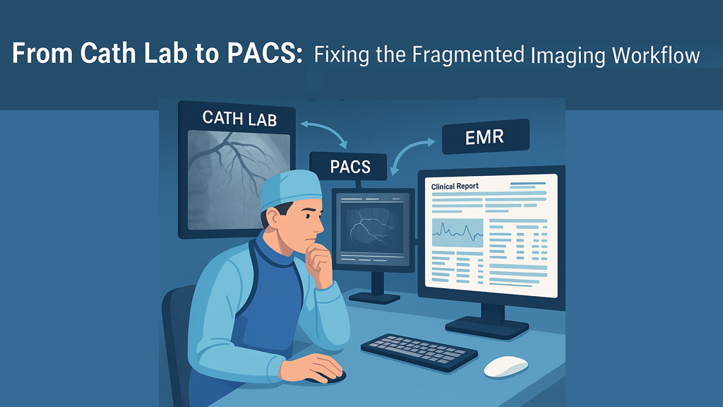
Take the example of Dr. Nair. After completing a complex bifurcation stenting, she logs into her PACS to review the images. But the patient’s lab reports are stored in a separate EMR system. The discharge notes are on a third platform. If she wants to correlate imaging with medication or lab trends, it involves manual cross-referencing, screenshots and a lot of wasted time.
This isn’t just an inconvenience — it’s a workflow bottleneck that affects:
- Clinical decision-making
- Interdisciplinary collaboration
- Documentation speed
- Patient follow-up quality
Where Things Break Down
The problem lies in how cath labs, PACS, EMRs and reporting systems were never truly designed to work together. Over time, institutions have added more tools without centralizing data access. This leads to:
- Redundant data entry across platforms
- Difficulty locating prior images or notes
- Delayed reporting and reduced case throughput
- Challenges in presenting cases for review or teaching
Why Centralized Imaging + Report Access Matters
Imagine being able to access a patient’s angiogram, ECG report, and clinical summary — all from the same interface. For busy cardiologists managing 20+ patients a day, this kind of integration would:
- Reduce cognitive overload
- Improve diagnostic accuracy
- Simplify multi-disciplinary case reviews
- Enable faster decisions in emergency scenarios
Fortunately, some modern solutions are moving toward unified systems that bridge these workflow gaps. Tools now exist that combine DICOM viewing with case summaries, patient notes and export options — all without requiring installation or complex IT infrastructure.
👉 Explore how efficient, portable DICOM management is shaping smarter workflows
Real-World Fix: A Teaching Hospital’s Experience
One tertiary care center in Pune transitioned to using a compact, portable system that allowed cardiologists to carry patient imaging + reports across departments. Not only did it speed up morning rounds, but it also empowered junior doctors to review cases more independently, without hunting for fragmented files.
“We didn’t need to revamp our entire IT infrastructure — just connect the dots more efficiently,” shared the hospital’s interventional chief.
Tips to Improve Imaging Workflows
- Audit your current workflow: How many platforms do you log into daily?
- Encourage tools that centralize access rather than add new silos.
- Prioritize portability — newer devices now offer plug-and-play access to DICOM + reports on the go.
- Look for formats that export images and summaries together (e.g., JPEG/AVI + case details).
- Collaborate with IT to reduce viewer login friction across devices and locations.
💬 Food for Thought
🔹 Are your imaging and clinical workflows connected — or coexisting in isolation?
🔹 What would a truly integrated system mean for your efficiency and patient care?
🔹 Have you explored newer portable tools that merge DICOM viewing with clinical reporting?
Efficiency in the cath lab doesn’t end when the procedure does. By closing the gaps between imaging, reporting and decision-making, clinicians can focus more on what they do best — treating patients.
If you’re curious about how portable tools are helping cardiologists manage cases more fluidly, take a look at this feature breakdown of what’s becoming possible.
0 Comments